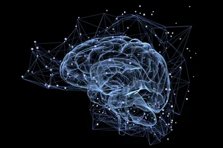
A remarkable paper just out in Nature has revealed images of the brain's structure and function in unprecedented detail: Network anatomy and in vivo physiology of visual cortical neurons.
Harvard Medical School researchers Bock et al took a mouse - just one - and used two forms of microscopy to investigate a small patch of it's primary visual cortex, the area which receives input from the eyes.
First, they used two-photon calcium imaging to look at the functional properties of individual cells. They displayed various kinds of patterns in front of the mouse's eyes, and looked to see which cells lit up, using a special dye which become fluorescent in the presence of calcium, which rises inside cells when they fire.
Having done that they took the same chunk of cortex (a rough cube of about 0.4 mm on each side) and used electron microscopy to see it in its ...


