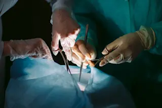
Livers outnumber people in Catherine Kling’s operating room at the University of Washington Medical Center. On this particular day, the extra organ — the only one ex-vivo, cleaned and sitting on ice — arrived just hours before the transplantation, the culmination of a thoughtful and time-consuming process of diagnoses, donor locating, evaluations and transportation, all sequenced by many expert pairs of eyes and hands.
Standing over the operating table as she’s done hundreds of times before, Kling, a general surgeon and transplant specialist, surveys the open belly in front of her. She quickly identifies the malfunctioning liver’s four main structures: the hepatic vein, portal vein, hepatic artery and bile ducts. After a few hours spent separating connections from the vena cava and nearby tissue, the patient's liver is fully removed. Next, Kling first attaches the replacement liver’s two main veins before removing the clamps and allowing it to fill with ...


