We have completed maintenance on DiscoverMagazine.com and action may be required on your account. Learn More

The Crack Team That Removes & Preserves People's Brains Just Hours After They Die

An Arizona retirement community helps create the world’s greatest brain bank. 


By Jeff Wheelwright
Oct 16, 2012 5:00 AMNov 12, 2019 5:26 AM
brain.jpg
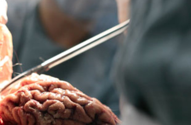
Doctors recover tissue during an autopsy at the institute in Sun City. | Courtesy Banner Sun Health Research Institute

Newsletter

Sign up for our email newsletter for the latest science news
 

On average, the residents of Sun City, Arizona, occupy their domiciles for a dozen years. When they depart—almost always by dying—they often leave their brains behind. The stages of physical and mental decline take them from their dream house to a hospital off Del Webb Boulevard, then to a nursing home, and finally back to the medical complex, where researchers harvest their most important organ. Hoping to do good for science, they have enrolled in the Brain and Body Donation Program of the Banner Sun Health Research Institute—widely considered the world’s preeminent brain bank.

A large base of well-
documented donors in close proximity sets the Sun City program apart from other repositories, which often have scant information about patients who may be scattered and diverse. Here, healthy, active seniors who eventually die of, say, heart disease, can be compared with others who develop neurodegenerative disorders. Because the two sets of subjects have similar backgrounds, lifestyles, and ethnic traits, changes relating to a brain disease should be easier to detect.

The institute is also famed for its crack autopsy team, which responds so quickly that no more than three hours elapse from the time a donor expires to the time that the brain is removed and preserved. “We’re not the biggest brain bank in the world, but we have the highest-quality tissue,” says pathologist Thomas Beach, the program director, who notes that donors must live within a 50-mile radius of the morgue.

After withdrawing some blood and cerebrospinal fluid for analysis, a team of rotating techs on duty 24 hours a day remove the top of the skull and take out the brain. The next step relies on a device that resembles a bread slicer, which is used to cut the brain into sections one centimeter thick. The slices from the left side are fixed in formaldehyde and those from the right are frozen between sheets of dry ice. Part of the tissue from the formaldehyde sections is stained, pressed into slides, and put under the microscope to verify the brain’s condition, healthy or diseased. The rest, light brown and convoluted, may be held in Tupperware containers at the research institute indefinitely. A recent visit to the storeroom turned up a container dated 1994.

These fresh brain sections, kept in carefully monitored freezers, are hot properties for advanced neuroscience research. Because the brain’s proteins, DNA, and other molecules are still intact, pharmaceutical companies are willing to pay high prices for the tissue. One of the most prized specimens is a sample of the entorhinal cortex, regarded as the X on the brain’s treasure map by Alzheimer’s researchers because the disease is thought to originate there; the nonprofit brain bank charges up to $1,000 for half a gram.

“Alzheimer’s starts at least 30 years before it’s diagnosed,” says Banner Health neurobiologist Paul Coleman, who works with entorhinal cortex from the brain bank himself. “The clinical trials for treatments have all failed because by the time of diagnosis the brain is so far gone that it’s like pouring gas into a car that has no engine.” Tissue samples from the ostensibly healthy brains in the institute’s brain bank might contain the seeds of the disease and clues to its treatment.

The brain bank has provided raw material to 110 investigators and several hundred studies over the past five years. Asked to name the most important use of the samples so far, Beach thinks for a moment and then describes an ambitious gene-expression study, “the first thorough study of gene expression of individually selected nerve cells in several regions of the Alzheimer’s brain.” And, he adds, “it’s publicly available.” Another project enabled the first FDA-approved imaging agent that could be used in PET scans of Alzheimer’s patients who were still alive.

In 2005 the institute expanded its autopsy program to include body as well as brain donations. The goal is to correlate the neurological changes found in Alzheimer’s or Parkinson’s patients with biomarkers of brain diseases in other organs. Scientists using Sun City tissue were the first to thoroughly map Parkinson’s lesions throughout the body and brain.

1 free article left
Want More? Get unlimited access for as low as $1.99/month

Already a subscriber?

Register or Log In

1 free articleSubscribe
Discover Magazine Logo
Want more?

Keep reading for as low as $1.99!

Subscribe

Already a subscriber?

Register or Log In

More From Discover
Recommendations From Our Store
Shop Now
Stay Curious
Join
Our List

Sign up for our weekly science updates.

 
Subscribe
To The Magazine

Save up to 40% off the cover price when you subscribe to Discover magazine.

Copyright © 2024 Kalmbach Media Co.