55-year-old Ruth Pavelko lies in an operating suite that looks a bit like the mission control center for a rocket launch. In the darkened room, physicians and nurses gaze up intently at glowing monitors as interventional cardiologist Emerson Perin injects stem cells into Pavelko’s weakened heart.
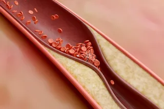
Pavelko, who has diabetes, wasn’t even 50 when a blockage cut off blood to part of her heart muscle. By the time she was 55, she had suffered three more heart attacks, and despite 13 stents to prop open her arteries, the damage has caused her left ventricle to balloon in size.
Patients with extremely advanced heart failure may become eligible for an assistive device, an artificial heart, or a transplant. But a clinical trial at the Texas Heart Institute in Houston provided Pavelko with another option: injections of stem cells extracted from her own bone marrow. These progenitor cells can differentiate into a few types of cells, including those that form blood vessels.
Looking at the monitors the day of her operation, it is easy to see why Pavelko’s heart is failing. On one screen a fluoroscopy scan monitor, or moving X-ray, shows her heart’s lopsided pumping. Another monitor transmits a color-coded image Perin has created using electromechanical mapping technology that was originally devised to track missiles. Some of the heart muscle courses with blood, but other areas are deserts of dead tissue without blood vessels. The 3-D mapping technology allows Perin to determine in real time the viability of cardiac tissue and decide which areas are hospitable to stem cells. The distinction is vital; injecting stem cells into scar tissue could create more scar tissue.
Perin has introduced a catheter into Pavelko’s heart through a needle puncture in her groin. When the sensor on the tip of the catheter touches the wall of her contracting ventricle, a computer follows the pitching of the sensor as it rides the contraction and assigns a number indicating the degree of the motion’s abnormality. Meanwhile, electrodes on the tip read whether the tissue is conducting electricity and therefore how well the muscle there can contract. Perin has mapped about 70 spots on Pavelko’s left ventricle. Although some spots aren’t contracting well, the readings show they are still viable and could be receptive to stem cells.
“Here we go,” Perin says, his eyes scanning the monitors. When he pushes the sensor against the inside of a ventricle wall, the computer reveals—and a tech calls out—numbers indicating that the wall’s movement is impaired, but its voltage is fine. Perin’s research assistant, Guilherme Silva, establishes that the needle is appropriately positioned to inject stem cells. “Injecting . . . injection complete,” Silva calls out, as if the first stage of a rocket has finished its burn and the second is igniting.
When this operation is completed, about 30 million cells—one million of them stem cells—will have found their way into Pavelko’s heart tissue. She is one of 16 patients in the first U.S. trial using cells extracted from adult bone marrow to reverse advanced heart failure. If the treatment succeeds, more than 5 million Americans could eventually be candidates for the procedure.
The first trial of adult stem cells for the heart, in 2000, used a type of lesser-known stem cell garnered from thigh muscle, not bone marrow. Philippe Menasche of the Georges Pompidou European Hospital in Paris infused the cells into patients. At first their heart function seemed to improve, then many patients developed arrhythmias, presumably because thigh muscle contracts differently from heart muscle.
A year later, Donald Orlic, a National Institutes of Health staff scientist, reported that stem cells derived from bone marrow and injected into damaged mouse hearts improved function in 68 percent of compromised areas. Although many scientists doubt that Orlic’s bone-marrow stem cells actually morphed directly into new heart tissue, they do believe the cells may have secreted powerful chemical cues—growth factors—or perhaps changed into blood vessels that revitalized the hearts.
Perin began experimenting with electromechanical imaging in 2001. He had planned to use this imaging technique to deliver growth factors to ailing hearts. When he heard about Orlic’s bone-marrow experiments, he realized that electromechanical imaging might improve the ability to deliver stem cells to hospitable tissue.
Many scientists, including some at the Food and Drug Administration, worried that clinical trials were premature. A few studies showed that stem cells from bone marrow could create scar tissue. Other studies hinted that stem cells, when released into the bloodstream, could get caught in narrowed vessels and clog them further. “It can depend on how you are looking at it, whether stem cells are good or bad,” says NIH cardiologist and stem cell researcher Richard Cannon. Perin says it’s easier to be cautious as a scientist than as a physician. “They don’t have patients dying; I do. Daily I have to look them in the eye.”
Perin decided the safest approach would be to inject the cells directly into scarless muscle, where they couldn’t get confused by either disordered chemical signals from inflamed tissue or a lack of signals from dead tissue. The imaging technique would allow him to do that. By the end of 2001, he was ready. He decided to launch his trial in Rio de Janeiro, where he had a hospital affiliation.
The trial was risky. No results from trials involving stem cells and multiple heart attack patients had yet been published. And his patients were very ill. One was so sick, Perin remembers, that “he was ashen, unable to breathe, starving—you can’t eat if you can’t breathe. Ejection fraction [pumping ability] was 10 percent. Under that you’re dead. Normal is 55. I found a viable area, but the rest of the heart looked dead. I was really worried.” An early death could end the trial. “I was sweating bullets. I took a deep breath, injected him, went to Texas. When I came back in four months, there he was, tan and smiling. In a month, he was a beach jogger.”
Before long, 13 of 14 patients given the procedure were able to trap and use oxygen better, an important determination of whether patients belong on heart transplant lists. Four of five Perin patients who had been awaiting a heart transplant were removed from the list.
The paper describing the Brazilian trial was published in the top heart journal, Circulation, in May 2003. By then, a few European papers were also revealing clues that stem cells derived from bone marrow and shot into the bloodstream of recent heart attack patients produced positive results. “Virtually everyone has reported some benefit,” says Cannon. But Perin was the first to show that the cells could restore function to far sicker hearts. And he had done it with an approach that released only a few cells into the bloodstream, thus avoiding the danger of clogging arteries. In March last year, the FDA approved Perin’s request to conduct the country’s first advanced, randomized, double-blind clinical trial of stem cells for heart failure. Perin will not have official results about Pavelko and others until next year, but he is already preparing to begin a clinical trial in Spain this year.
Still, no one knows. The race is on not only to prove that patients are getting better—by comparing patients treated with stem cells with untreated patients in a blinded trial—but also to pinpoint the effects. Most researchers believe that bone-marrow stem cells prompt new blood vessels—not heart muscle—to form. One way to know is to study stem cell injections in pigs, whose hearts are similar to humans’ in both size and physiology.
In a study of 32 pigs, Perin’s team has been looking at the effects of injecting varying quantities of bone-marrow stem cells into the heart. The group is also testing four other types of stem cells. The first step in the experiment is to induce a condition preceding heart failure. The researchers slip a band filled with gel around a pig’s heart artery. As the gel slowly absorbs fluid, it constricts the artery until it is almost or totally blocked. A month later, the pig receives its allotted injection of bone-marrow stem cells.
One month after the injection, a researcher takes moving X-rays to see how the ventricle is doing. An injection of dye floods the pig’s branching coronary arteries like lightning setting off a gnarled oak on a dark night. Perin also uses his imaging technology to get a more precise view of cardiac function. He draws the catheter and aims at 66 positions, with the technician locking in the electrical and mechanical coordinates each time. When he is done, he examines the before-and-after shots on a monitor. In this case, areas that were dying a month ago have shrunk and healthy areas have enlarged. “Before, voltage on the anterolateral wall was 7.6,” says the technician. “Now it’s above normal: 18.4. Drastic improvement.”
“Beautiful,” Perin says.
Hours later, he relaxes in his office, which overlooks the Texas Medical Center, the world’s largest such complex, where the first successful U.S. heart transplants were performed in the 1960s. His optimism about stem cells and heart disease is unquenchable, and he talks up another project—to use stem cells to wean dying patients off heart machines.
Other researchers see the potential as well. Doug Losordo of Tufts University is heading clinical trials in Massachusetts, Minnesota, and California to test a subset of the cells Perin uses. Studies suggest this more specialized cell type may prompt better blood-vessel formation. The technique carries the risk of clogging arteries because a drug is injected that forces swarms of stem cells out of the patient’s bone marrow and into the blood, where they circulate for days before being collected for injection into the heart. But Losordo’s patients wear vests that monitor and control arrhythmia, and so far there have not been problems. Because his patients are healthier than Perin’s, and are getting concentrated shots, they may experience even better results. Losordo says most of his treated patients “have had symptomatic improvement.”
Amit Patel, a cardiac surgeon and researcher at the University of Pittsburgh, has completed three clinical trials in Asia and South America that included more than 100 heart-failure patients. He used cells similar to Losordo’s, and he says his patients’ hearts have been pumping much better. In one trial, a measurement of pumping ability almost doubled, going from 26 to 46—nearing the normal of 55 percent. Much of the improvement Patel has seen, he says, is “unbelievable.” Two papers are in the works, and in May the FDA approved a U.S. trial. Johns Hopkins is also starting a clinical trial using stem cells in heart attack patients.
Perin welcomes the studies. The faster the FDA moves, the more doctors can look patients in the eye. “That’s what matters,” he says.
The best proof of success will come from dissecting hearts—which can’t be done in living people. The death of a 57-year-old patient in the Brazilian trial from an unrelated stroke 11 months after treatment uncovered a long-awaited clue. Perin calls up two images on his computer: “Look,” he says excitedly.
The pictures resemble shots of multicolored stars. On the left, a few stars; on the right, a small galaxy. The left image is the back of the ventricle, where no cells were injected. The right image shows the front of the ventricle, where Perin injected 30 million cells. Glowing antibodies identify substances found only in growing cells. The area where the cells were injected blooms with the markers.
Perin also used antibodies to label cells expressing vimentin and desmin, proteins found on developing, not mature, heart muscle cells. Only blood vessels and muscle in the section injected with stem cells lit up. “You don’t have to be a rocket scientist to get this,” he says. “Vimentin and desmin shouldn’t be there. But they are. When I show this to cardiologists, they say, ‘This is not normal.’ We can’t know if these are old cells rejuvenated by transplanted stem cells or new cells maturing from them. But it’s undeniable. Something new went on in this guy’s heart.” The journal Circulation agreed and published Perin’s paper describing the results in July.
Ruth Pavelko doesn’t read cardiac journals, but she knows something is different in her heart. Before she received her stem cell injections last January, she could not walk to her mailbox. She could not climb the stairs to her bedroom without help, and even the lightest chores were taxing. Today she climbs up and down stairs many times a day, does laundry and dishes, and once a week does a quarter-mile jaunt around a nearby park. “I no longer have that heavy feeling on my chest, like someone is sitting on it.”


