We have completed maintenance on DiscoverMagazine.com and action may be required on your account. Learn More

Inside the Human Brain

With the power of neuroimaging, David Ewing Duncan takes a magical mystery tour of his own mind, exploring fear, memory, and faith.

By David Ewing Duncan
Mar 25, 2019 4:45 PMNov 12, 2019 6:24 AM
ScreenShot20140814at25854PM.png
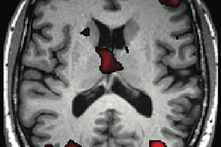
An MRI scan of Duncan’s brain highlights areas (in red) that screen out irrelevant information—a process that weakens as we age. 

Newsletter

Sign up for our email newsletter for the latest science news
 
Author David Ewing Duncan wearing an EEG cap.

James Brewer takes a seat beside me in a cafe at the San Diego Convention Center, where we are both attending the largest neuroscience meeting in the world: thirty thousand brains researching brains. With his balding head, bright eyes, and baby cheeks, Brewer, a neurologist at the University of California at San Diego, looks like a large and curious toddler. An unlikely messenger, perhaps, in what for me is a moment of truth. I had undergone a series of diagnostic procedures in his laboratory, and now, inside the laptop he has placed on the table, are the results of my brain tests.

“Your brain is shrinking,” he says.

This is the last thing I expected to hear. Not me, a man who considers himself healthy and ageless, at least in his own, er, mind.

“People’s brains begin to shrink when they are in their thirties,” Brewer explains with a smile, to suggest this isn’t really a big deal. “Yours is about average.” I’m somewhat reassured but still concerned about what else I will soon learn. Brewer’s tests are just the first stage of a thorough investigation to see what state-of-the-art medical technology can tell me about the health of my noggin, part of a project I’m calling Experimental Man. I am exploring what diseases might be lurking in my head; what my memory is like at the age of 51; and how my brain responds to matters as diverse as fear, greed, the movies I like, and even the idea of God. It is a magical mystery tour of a single person’s brain. Mine.

Within the hundred billion neurons and trillion or so glial cells that hold neurons in place and feed and protect them lie my hopes and fears, my feelings, my memories, even the words now recorded on this page. Without my specific blend of cells, tissues, genes, and experiences, there would be no individual person to take these tests, no personality to try to understand what the results mean.

Every brain contains secrets: memories we would rather dispose of, proclivities that we are not proud of or that we struggle to control. For instance, when I was younger I had an almost debilitating anxiety about certain social situations, a predilection I have now mostly learned to control. Yet the fear remains that I will say the wrong thing or do something embarrassing in public. I sometimes wonder if my anxieties are normal—something that through this investigation I would be able to find out.

Before I launch this journey into my brain’s subtle workings, though, Brewer is taking a look at the overall structure of my thinking machine. I have already taken a “structural” MRI (magnetic resonance imaging) scan, which has evaluated the size and well-being of anatomical features in my brain, creating a map Brewer is about to show me on his laptop. Now I will see what these structures, such as the cerebellum and hippocampus, look like. He tells me that abnormalities in the brain can offer evidence of diseases that are present now or that may strike in the future. I already know, for instance, that patients with schizophrenia have distinctive aberrations in the right superior temporal gyrus and that those with attention-deficit/hyperactivity disorder have smaller brain volumes in all regions.

The brain images Brewer shows me —views from the side, back, and top—look like a cross between a nice, fat slice of sausage and a Rorschach inkblot test. A variety of computer- generated colors highlight my hippocampus, lateral ventricles, and other features that Brewer has measured to determine whether I have early signs of diseases such as Alzheimer’s. 

Brewer calls up a picture of the brain of a person who actu- ally has Alzheimer’s, and I see that various anatomical features look as if they are smushed or blurred. This distortion results from atrophy and an overaccumulation of proteins that cause the hippocampus to shrink and adjacent fluid-filled structures to expand into the space where the rest of the hippocampus used to be. Fortunately, my hippocampus seems fine. The only abnormality is a slightly larger left brain, the seat of most of what happens when we use language. “But as a writer, you didn’t need a scan to know that,” Brewer says. He adds, however, that he is mostly kidding. There is no hard evidence that the size of the left or right hemisphere affects one’s personality, despite pop neurology assertions.

Weeks later in another research lab, I will be scanned for brain function: what goes on in my brain when I experience fear or religious faith or when I make decisions. A functional MRI scan, known as an fMRI, will check for increases in blood flow in certain areas of the brain as I react to or record an experience, initiate an action, or ponder something. Blood is needed to fuel the neurons when they go to work, and the fMRI scan picks up signatures of the iron in the blood. From this information a neural map can be created showing where brain activity occurs and how much blood is flowing there.

Since MRIs began to appear in the 1970s and 1980s in labs across the country, neuroscientists have been running virtually every fMRI scan imaginable, checking brain reactions to everything from pornography to pictures of sunsets. Nevertheless, applying MRI technology to healthy individuals to ascertain their proclivities toward certain diseases or behaviors is considered to be in its infancy. “These tests are so early, they are almost totally irrelevant to individuals,” warns Judy Illes, a neuroethicist at the University of British Columbia, “though I think there is so much that seems tantalizingly close to being understood.” At the National Institute of Neurological Disorders and Stroke (NINDS), neurologist Eric Wassermann agrees. “Data for brain scanning are noisy for an individual,” he says. “What we have is mostly group data relevant to, say, an age group or to people with or without a brain disorder.”

Yet Illes and Wassermann are enthusiastic about my inves- tigation, seeing it as an opportunity for a nonscientist to describe what, if anything, MRIs and other emerging brain- reading technologies can tell us about the three pounds of mushy tissue between our ears, home organ to that mysteri- ous realm called the mind. 

Memory

On a perfect Bay Area day, with the temperature in the upper 60s and a hazy, warm sun shining, I’m in a brain lab at the University of California at San Francisco, attached to a machine that is going to test my ability to remember. Students of UCSF neuroscientist Adam Gazzaley have applied a gooey gel to my head and strapped on an electrode monitor, which looks like a cross between a bathing cap and a device a mad scientist might use to zap my brain. The tight plastic cap is covered with dozens of electrodes connected by wires to an electroencephalograph (EEG), which measures electrical activity in the brain.

Through his research, Gazzaley has set out to prove that the brain does not lose its ability to remember as it ages but rather loses its ability to filter out unwanted memories; in a sense it remembers too much. He is demonstrating that the frontal lobe—the rational and decision-making part of the brain—has a major influence on what is retained by the hippocampus and the brain’s other emotional centers. Neuroscientists call this function top-down modulation, a process by which the frontal lobe guides both what people recall and what they do not want or need to burn into their memory cells. The brain does the same thing with hearing. Young ears (and brains) can easily discern the voice of a friend sitting across a table in a noisy bar; as ears and brains age, they are less able to pick out the friend’s voice from the barrage of other sounds. “We think it’s a filtering problem,” Gazzaley says. 

As the students strap the cap on me, Gazzaley explains that I’m an anomaly for this experiment. At age 51, I fall in between his two test groups: the young’uns, ages 19 to 30, and the seniors, ages 60 to 77. (I am actually much closer to the older group, but who’s counting?) This makes me wonder, as they ask me to fit my chin into a viselike contraption that will hold my head still: Will I test closer to the young subjects or the old? The experiment has subjects look at numerous photographs of faces and landscapes shown for one second each on a monitor. Then the screen goes blank for eight seconds, after which participants are shown either a face or a landscape and asked, “Was this one of the images you just saw?” I’m told to click on a box I’m holding in my hands: yes with my left thumb, no with my right. To test the idea of filter- ing, Gazzaley asks subjects in some of the tests to ignore the faces; in others, to ignore the landscapes. He also runs series of photographs that are only passively viewed, to ascertain a baseline of what lights up in the brain when a person is not actively trying to suppress or enhance faces or landscapes. 

I am feeling a touch of performance anxiety. James Brewer has already knocked down my sense of agelessness a notch or two with the news that my brain is shrinking. Now I am subjecting myself to a test that might reveal me to be prematurely transforming into mental codger-hood. 

With students in another room monitoring my responses, I’m alone in the dark, staring at a computer screen a few feet in front of my face. Being the competitive sort, I am highly motivated to get the right answers, and I get mildly ticked off when I miss one. As the test wears on, however, I begin to get sleepy—a big problem for excelling at what is essentially a boring, repetitive task. 

Afterward I wash the goo out of my hair and join Gazzaley and his students to review my data. My thought waves appear as jerky lines running across a monitor, like multiple tracings from a polygraph test. 

Am I OK?” I ask, slightly nervous as I realize that this EEG is what amounts to my brain talking in streams of electricity.

“We’ll see,” Gazzaley says. 

Later that day, the team repeats the same basic experiment with the faces and landscapes, except this time an MRI measures the blood flow in my brain as I react. 

A few weeks later, Gazzaley summarizes his findings and tells me he sees nothing alarming or unusual in my results for memory. “You aren’t abnormal, and you have no evidence of disease that affects your memory,” he says, pulling up slides of my results on a large computer screen on his desk. “You did great on reaction times and accuracy, better than even the younger group.” This is unexpected, given that I was close to falling asleep for part of the experiment.

I am pleased by this—it almost mitigates the knowledge that my brain is shrinking—but then Gazzaley reveals that I did not do as well on filtering the distractors. “You came out where we expect you to be at your age, about halfway between the younger and the older groups.” Like the older group, I remembered things fine, but my brain betrayed me by revealing that I was not always filtering out what I was supposed to ignore. More precisely, my aging brain is unable to suppress irrelevant information either as well or as quickly as when I was younger.

“It means your brain is aging,” he says. “Sorry about that.”

An MRI scan of Duncan’s brain highlights areas (in red) that screen out irrelevant information—a process that weakens as we age. 

High Anxiety

Deep in the tunnel of another MRI machine, this one at Stanford University, I am reliving one of the most anxious moments of my professional life. 

It happened years earlier when I was a junior correspondent for Life magazine. I was in a staff meeting led by managing editor Dan Okrent, a legendary and (to me) intimidating veteran of books and magazines. We were discussing one of the first major stories I reported, a possible cover piece. Colleagues were saying I did a great job and that the photographs shot by Joe McNally were fantastic. In those days I was quite shy in such meetings with senior editors, but I was feeling good. Then Okrent, who could be gruff and blunt, blurted out that another reporter would write the story and get the byline. I couldn’t believe it. As the meeting continued, I felt my heart rate surging and my gut contracting. I felt ashamed, and I am sure my face turned red. I knew I should say something. I needed to stick up for myself, but my overriding desire was to say nothing. I had to regain control. 

Inside the MRI machine, the intensity of the emotions from that long-ago afternoon are flooding back into my brain as I hear the distant whir of the great magnet wrapped around my head and a stream of clicks and sounds of grating metal. On a monitor above my eyes, the tale of the Life magazine meeting is being displayed in reminiscences that I have written at the request of Philippe Goldin, a Stanford research scientist, and Kelly Werner, a postdoc, both working in the lab of Stanford psychologist James Gross. In their work Goldin and Werner ask for stories that are personal and socially embarrassing and that seriously punish their subjects’ self-esteem. They then use fMRI scans to investigate the phenomenon of social anxiety and examine those who suffer from phobias and disorders that make them terrified of social situations and personal interactions. 

Fear and anxiety are crucial to human life. They are evolutionary tools developed over the eons to protect us. They alert us to run for the hills when a lion roars and to fret when our children are sick so that we will take care of them. My genetic forebears, however, never had to face a humiliation delivered by the managing editor of Life magazine. 

Despite my nervousness about this test of nervousness, at least I am being examined as a healthy subject—I hope. Researchers have found that the imaging of supposedly normal brains reveals clinically significant findings 8 to 10 percent of the time—“disconcertingly often,” says Henry Greely, a law professor at Stanford who works on legal and bioethical issues that include neuroscience, genetics, and stem cells. 

In the scanner, the Life magazine story continues to flash in front of my eyes. I’m supposed to rate how anxious the statements make me, on a scale of one to five, by punching the appropriate button on the response pad in my right hand. 

Back in New York, the managing editor at the weekly staff meeting congratulated me—and then assigned the story to a veteran reporter to write. 

I press four, meaning this memory still makes me pretty darn anxious. The screen clears, and up pops another sentence in bold capital letters, this one written by the researchers: I am a loser. 

This is intended to induce “negative self-beliefs” to see what happens in my brain. “Some people launch into a cascade of negative self-beliefs,” Goldin says. “We want to track how that happens.” 

I am too insecure to stand up for myself.

The test then asks me to make an effort to modulate these negative self-beliefs, to use strategies to tell myself I’m not that bad. This is the second part of the experiment: seeing how well people’s strategies work to alleviate their anxiety. Once I have made an effort to chill out, I am again supposed to press a button to indicate how I feel. 

People will think I’m a wimp.

“The amazing thing is that people’s behavior can make changes in the brain,” Goldin says. “Cognitive behavioral therapy—the gold standard of psychotherapy—and mindfulness meditation training both help to reduce activity in the amygdala. It can be tempered to learn and adapt.” 

In my Life magazine case, I overcame my terror and stuck up for myself after the meeting. Okrent agreed to let me write the next story that I reported. This experience was among the first of many where I worked hard to modulate my fears, apparently pushing my amygdala—one of the seats of emotion activated by social anxiety—to shape up. This largely successful battle to squelch my anxiety could be clearly seen in my scans. In one, my anxiety is shown as my brain’s amygdala lights up—along with the visual, color, word, and number recognition centers—as I relive a vivid emotional memory. A second scan shows areas of my brain associated with language and modifying beliefs flare up as I try to regulate my anxieties. 

“You seem to be able to control your emotions, which is good,” Goldin says. “Some people have exaggerated reactions.” I tell him I’m surprised at my ability to tamp down my reactions, given my state of high anxiety when I was younger. “You’ve been alive long enough to learn to use your mind to respond skillfully to anxiety,” he answers. “That’s what people are supposed to do.” Still, as I write this, I’m looking around at people in my favorite café near my home in San Francisco and wondering: Are some of them even more anxious than I am? Or is my brain just better at covering it up? 

Religion and Baseball

Now I’m in Bethesda, Maryland. Another day, another MRI scan. This time, the prompt on the monitor I’m gazing at inside the machine leaves no room for a nuanced answer: There is a god. 

I have a few seconds to answer yes or no on the clicker in my hand, but I am stumped about which button to push. 

As blood surges in my head to locales associated with religious belief, I’m thinking that this question, for me, may be unanswerable. I am essentially nonreligious. I seldom go to church, and I often find myself agreeing with the likes of Sam Harris and Christopher Hitchens that organized religions are mostly artifacts of premodern cultures that in ancient times created all-powerful deities to explain and cope with the unknown. I believe that overzealous piety has led to horrors such as the Inquisition and to dogma that at times becomes so rigid that it blatantly contradicts scientific proof (and sometimes common sense). Yet I know that religion clearly comforts people. Studies show that patients who pray often tend to do better than those who do not. Nor can I deny the crucial importance of spirituality, a sense that one’s goals can be bigger than just looking out for oneself. 

Bottom line: I have no proof that a god exists or that the universe is anything but random atoms assembling and disassembling without a design or a creator. My thumb twitches above the “no” button. 

And yet I lack definitive proof that God does not exist. It is possible that he (or she or it) is real. Not the man with the beard depicted in medieval paintings, but some force far beyond our brains’ comprehension. If there is even a 0.0001 percent chance that this is so, can I answer no? Time is up. My thumb moves toward yes, and I press it. I feel exhausted by so many thoughts racing through my brain; the neuronal exertion must have lit it up like a city at night seen from 35,000 feet. 

That was exactly the point of the test, says Dimitrios Kapogiannis, a neurologist at NINDS. Kapogiannis and a small team of researchers asked 40 subjects these same questions about God and religious beliefs while they were in the MRI machine: 20 who said they were religiously inclined and 20 who said they were not. “The purpose of the study has been to discover the underlying cognitive structure of religious beliefs—to find out what cognitive processes take place when religious and nonreligious people think about religion,” Kapogiannis tells me. “Then we want to identify brain regions that become active with each such process.” 

For this experiment Kapogiannis is working with cognitive neuroscientist Jordan Grafman. Chief of the Cognitive Neuroscience Section at NINDS, he is a researcher with a longtime interest in trying to understand how the brain works when people are identifying with cultural phenomena and beliefs. In 2006 Grafman’s lab ran an experiment on political beliefs, scanning the brains of people as they viewed pictures of John F. Kennedy, Hillary Clinton, Ronald Reagan, and John McCain. Grafman’s team found patterns of blood flow in specific areas of the brain suggesting certain ideological proclivities that could be correlated with party affiliation. “The core of the brain is conservative,” Grafman says, but “the outer shell is liberal; it’s playing with things, trying things out.”

A functional MRI scan measures blood flow in Duncan’s brain while he challenges his memory. Colored areas show his motor cortex activating as he responds to a test by clicking a mouse. 

Grafman, an affable, gentle man with large eyes, is using his scanners to peek into brains as they wrestle with big questions such as politics and religion. He has also delved into the social neuroscience literature to understand why anyone would love the Chicago Cubs, a team that has not won a World Series in 100 years. Grafman says that Cubs worshippers are similar to religious adherents in their staunch belief that every “next year” will be the year. He says this hope springs from the prefrontal cortex, the region of the brain responsible for high-level cognitive activities such as planning, reasoning, establishing context—and, undoubtedly, inventing justifications for loving perennial losers in baseball.

Back in the machine, the questions are still coming. Kapogiannis asks me 70 in all, covering subject matter from God as being angry and wrathful to God as being loving, with a variety of topics in between. I answer no to the “God is angry” sort of questions. I also answer no to questions about mixing God with politics and social policy. Finally come questions asking whether God is loving and compassionate; I would like there to be a good God who is kind and caring and a heaven that virtuous people end up in. But I see no proof of this, so I answer no. 

Weeks later, Kapogiannis sends me my results along with pictures of my brain and his interpretations. First up is how my brain reacted to the idea that God is involved in my life and in the world. Kapogiannis explains that this image showed elevated activity in the right side of my brain in action-oriented areas. “God’s presence activates a part of your frontal lobe concerned with understanding actions,” he says. “You are actively imagining a God in action, visualizing an involved God, imagining a God willing to intervene.” 

“But I answered no to the questions about God’s involvement and willingness to intervene,” I protest. “I fundamentally don’t believe this—unless I’m lying to my brain, which imagines a God in action without my knowing it.” 

“This is just one interpretation,” Kapogiannis says.

I tell him about the barrage of thoughts racing through my head during the question about God. “Could it be that I’m just trying to decide which button to press?” I ask. 

“That could be happening too,” he agrees. “This is all tentative right now.” Other results showed my brain getting very active over the social policy questions —probably because I strongly object to mixing religion with such issues as abortion and homosexuality—and relatively quiet when I was asked about God’s being angry or loving. “These inquiries didn’t cause a strong activation,” Kapogiannis tells me. “This is a pattern seen in other people who are not particularly religious.” 

I find Kapogiannis’s work fascinating, and I have no doubt that his research will help us understand the mechanics of how religion and belief work in our brains. But so far the interpretations of my true state of mind by using blood flow to regions of my brain seem at best sketchy—which he admits—and at worst little more than a sophisticated version of reading tea leaves.

My head is one of millions around the world being inserted into MRI scanners and getting hooked up to EEGs and other devices. Researchers are running thousands of experiments on everything from what our brains look like when we are watching TV to what happens when we are in love or lust. 

The tests I took for my personal experiment provide a sampling of what is available for an individual, though, to be honest, I’m not sure how much they enlightened me about myself. I am pleased to know that I have no identifiable traces of disease. As for the rest, I got what researchers promised with this young technology when they told me it is not yet ready to test individual brains: a number of intriguing images and mostly impressionistic interpretations. 

“One problem is that many of these studies tend to be one-offs—they’re done by one laboratory and not repeated,” says Judy Illes, who warned me early on that the science’s level of sophistication for individuals was not high. She notes that there are few follow-up tests or attempts to replicate initial data or to run the same experiments on larger populations, although she expects this to change. “Neuroscience right now needs a meta-approach linking all of this together,” she says, a project to create not only a vast map of the brain but a schematic of pathways and how different regions connect. 

At UCLA, a project led by neurologist John Mazziotta and research neurologist Arthur Toga is attempting to do exactly what Illes suggests: create a comprehensive atlas that will provide a template for what is now known about brains—what they look like, how they vary, and how they function. Collaborating with researchers from Canada, Europe, Japan, and, in the United States, the University of Texas, the brain atlas team has scanned 450 “normal” brains and used hundreds of thousands of images taken of 7,000 people around the world to compile three-dimensional color maps of the brain. These maps, they say, will show everything from relative sizes of anatomical features to differences in brains associated with age, race, gender, educational background, genetic composition, and other distinguishing characteristics. Mazziotta and Toga’s study also includes cadaver brains, cut up into more than 2,500 microscopically thin slices and mounted on glass slides, then stained and digitally photographed. 

Layered over the anatomical maps will be brain functions such as memory, emotion, language, and speech. The finished atlas, Mazziotta says, will serve a purpose similar to what the Human Genome Project has done for geneticists, providing a detailed framework of the brain that researchers can use to perform experiments. 

Whether the brain atlas will one day lead us to a metaunderstanding of this most mysterious organ will not be known for decades. As I contemplate all of this sketchy knowledge about myself and the many hundreds of experiments I could run to find out even more, my head begins to ache. So I decide to run another experiment: to clear my mind of all thoughts, feelings, and emotions. I try this for several minutes, wishing I had paid more attention to a friend who attempted to teach me meditation. Nope. Doesn’t work. Certainly somewhere in my brain blood is flowing, lighting up something in that mysterious lump of gray matter that is me. 

1 free article left
Want More? Get unlimited access for as low as $1.99/month

Already a subscriber?

Register or Log In

1 free articleSubscribe
Discover Magazine Logo
Want more?

Keep reading for as low as $1.99!

Subscribe

Already a subscriber?

Register or Log In

More From Discover
Recommendations From Our Store
Shop Now
Stay Curious
Join
Our List

Sign up for our weekly science updates.

 
Subscribe
To The Magazine

Save up to 40% off the cover price when you subscribe to Discover magazine.

Copyright © 2024 Kalmbach Media Co.