How Do Scientists Measure Brain Activity?

The brain is constantly buzzing with electrical activity. Here's how scientists study the signals that may someday help explain the nature of consciousness itself.

By Conor Feehly
Mar 24, 2022 5:00 PM
EEG
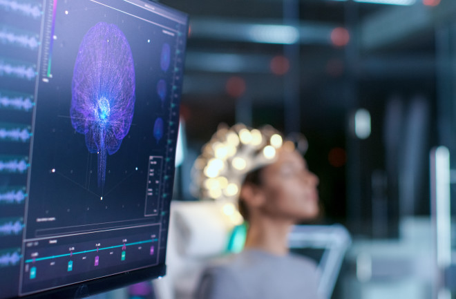
Electroencephalography (EEG) measures the collective firing of neurons across different neurological regions of the brain. (Credit: Gorodenkoff/Shutterstock)

Newsletter

Sign up for our email newsletter for the latest science news
 

What gives rise to our conscious experiences? The so-called hard problem, popularized by the philosopher David Chalmers in 1995, asks how the inanimate neural substrates of the brain create vivid first-person conscious experiences. In other words, how the combined firings of our neurons elicit the inner subjective universe we all possess. 

Anil Seth, one of the world’s leading neuroscientists and researchers of consciousness, says that scientists are gradually working to connect the dots between different aspects of consciousness and the dynamic states of the brain that give rise to them. And because any explanation that scientists provide for the inner workings of consciousness (and other cognitive functions) is attained by studying neural activity, it's important to understand how they obtain and analyze data on the brain.

At a basic level, electricity is the language of the brain. Continuously, electrical impulses, also known as action potentials, are buzzing around between your ears. Your neurons, and the synaptic junctions where neurons meet, are bathed in a chemical bath — in the form of neurotransmitters like dopamine — that mediate the electrical activity in their neighboring cells. This is the reductive basis of neurological communication, and somewhat mysteriously, these interactions underpin every thought, feeling, or action you have ever experienced.
But as scientists have learned more about the brain, they’ve noticed that large-scale states of brain activity are also important in its overall function. Different regions of the brain are known to synchronize as they fire neurons in tandem at different frequencies. These neural oscillations, or brain waves, are thought to facilitate communication between different regions of the brain. That means that scientists need to look at all scales of brain activity, both at the cellular level and within these larger networks, in grappling with the task of understanding consciousness: If we focus too closely on, say, the individual firing of neurons, then we risk missing the bigger picture, like mistaking a single drop of water for the entirety of the ocean.

Mapping Neural Activity  

One of the most widely-used methods for generating data on the brain while people carry out certain cognitive or behavioral tasks is functional magnetic resonance imaging, or fMRI. This non-invasive neuroimaging technique is a go-to for cognitive neuroscientists, largely because of the practical ease with which it can generate data. But fMRI doesn’t actually measure electrical activity in the brain; rather, it measures the indirect consequence of neural activity. Namely, where oxygenated blood is flowing.

This isn’t necessarily a problem. Thermometers don’t measure temperature directly — they measure the volume of mercury in a glass tube, which is tightly coupled with temperature. Problems can crop up when coupling is incomplete, noisy or complex. Meanwhile, blood oxygenation, also known as the hemoglobin response, is thought to be most tightly coupled with specific synaptic events that play a significant role in neurological communication. 

Much like a digital camera or computer screen, an fMRI brain scan can be defined in units of spatial resolution, but with images in 3D, rather than 2D. These volumetric pixels are called voxels; in a typical scan a voxel might cover 3 cubic millimeters of tissue, a volume that would carry approximately 630,000 neurons.

Few brain scanning methods have better spatial resolution than fMRI, barring intracranial recordings like electrocorticography (ECoG) that can isolate activity from single cells. But these invasive techniques, where electrodes are placed directly on the brain, are limited to animal models or specific clinical contexts where patients suffer from conditions like epilepsy that require a high level of precision when locating seizures. By contrast, fMRI strikes a good balance between precision and coverage. 

An fMRI scan performed during working memory tasks, which typically show activation in parts of the brain's frontal cortex and parietal cortex. (Credit: Public Domain/Wikimedia Commons)

A bigger problem for fMRI is that blood oxygenation is drawn out over time, which makes it difficult for neuroscientists to pinpoint precise moments of activity. Typical fMRI images are taken from an average of brain activity over time, usually just a matter of seconds, somewhat like a long-exposure photograph. And this can cause problems as highly dynamic or fast neural processes become blurred. 

Methods that measure electrical activity more directly give neuroscientists a better idea of when neurological events actually take place. Electroencephalography, or EEG, is another textbook way neuroscientists gather data on the brain. Whereas fMRI measures where oxygenated blood is flowing in the brain, EEG measures the collective firing of neurons across different neurological regions. One caveat with EEG is that it can be difficult to know where exactly the neural activity being measured is generated. Still, the method has been largely responsible for our understanding of neural oscillations, or the combined synchronized firing of neurons. If the brain was a stadium filled with people, and each person was a neuron, one part of the arena could communicate with another through synchronized activity, like clapping. 

Another common technique for neuroscientists to gather data on the brain is magnetoencephalography (MEG). Instead of measuring the electrical potentials themselves, MEG measures the tiny magnetic fields generated by the brain’s electrical activity. This technique involves a specialized helmet with around 300 extremely sensitive magnetometers that detect the very small magnetic fields produced by the brain. Such neural readings have to take place in shielding rooms, which block out the magnetic fields of outside objects — even the Earth’s magnetic field. 

Current MEG technology is limited to detecting magnetic fields in the cortex, the bit of the brain which is closest to the scalp. But there is still lots of interesting stuff happening in there, especially if you're investigating aspects of conscious experience, as the cortex is thought to be heavily involved in emotion, language, and memory. Magnetic fields generated in the deeper regions of the brain dissipate before they reach the surface of the head, where they can be picked up. It is thought that the magnetic fields measured in MEG are the result of the combined activity of at least 50,000 neurons simultaneously communicating with their neighboring neurons. If activity in the cortex is the subject of investigation, MEG provides the best data on when the activity took place and where the activity happened when compared to other techniques. 

Digging Into the Data 

Once scientists have collected data via these different technologies, it’s then up to them to decide how to analyze that data. A familiar result of brain imaging techniques like fMRI is to get a picture of the with different regions lit up, representing increased activity in those areas. 

In short, what these images show is contingent on analytic decisions made by scientists, and different types of analysis and sample sizes can yield dramatically different results. In a 2013 study, rather than relying on thresholds to determine activity and noise, the researchers determined anything that was found in multiple subjects could be considered a signal, and anything that didn’t would be considered noise. They found the same networks that are often mentioned in other research, but they also found that these networks overlap in regions called hubs, which are densely connected and are responsible for coordinating activity across the whole brain. 

Ultimately, the types of conclusions we can draw about the functioning of the brain, and how they give rise to conscious experiences, are limited by the types of data our technologies can provide — as well as the assumptions we bring to bear when analyzing that data. 

It’s fascinating to speculate about what types of dynamics may be unfolding in the brain that our current technologies aren’t capable of accessing. Neuroscientists are still working to develop new techniques for understanding the complex neurochemical interactions that take place between neurons, as well as attempting to map connections between different regions of the brain.

“In our field, techniques are constantly being developed or adapted to gather information on the dynamic properties of the brain,” says Robert Mason, a neuroscientist at Carnegie Mellon University, who describes two relatively new technologies in the field: TMS (transcranial magnetic stimulation) and tDCS (transcranial direct current stimulation). The basic premise behind the techniques, Mason says, is to stimulate the brain to determine how that impacts both behavior and the brain states being measured, with the potential to map out connections between different cortical regions. “Like most of the other brain measurement techniques, there are many underlying assumptions and it is not as simple as it first appears,” he adds. 

New techniques like TMS and tDCS could someday help scientists answer knotty questions about the nature of consciousness. Seth believes that the hard problem will slowly dissolve as we draw bridges between the characteristics of what it feels like to be human and their corresponding neurological processes. 

However, there’s also a chance that our techniques will be unable to provide us with a full enough picture of what is going on inside us to answer any questions about consciousness definitively. The near-incomprehensible complexity of our brains mean there are sure to be dynamic properties unfolding that scientists are completely unaware of. What’s more, we could even be missing something in the data that we already have. Still, it's not unfathomable that the secrets of the brain, which contribute to one of the biggest mysteries of them all, can one day be better understood. 

1 free article left
Want More? Get unlimited access for as low as $1.99/month

Already a subscriber?

Register or Log In

1 free articleSubscribe
Discover Magazine Logo
Want more?

Keep reading for as low as $1.99!

Subscribe

Already a subscriber?

Register or Log In

More From Discover
Recommendations From Our Store
Shop Now
Stay Curious
Join
Our List

Sign up for our weekly science updates.

 
Subscribe
To The Magazine

Save up to 40% off the cover price when you subscribe to Discover magazine.

Copyright © 2024 Kalmbach Media Co.