A Stanford Medicine team proposes that the brain is made up of two organs with separate origins. That distinction could help explain why some nerve cells, including those that help us swallow, have been so difficult to grow in a lab.
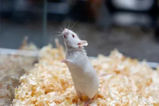
By studying mouse embryos, the researchers found that the cells forming the back of the brain develop separately from those forming its front and middle.
The findings, published in Nature Neuroscience, helped the team grow human nerve cells that send instructions to muscles involved in swallowing. Those cells could help researchers study ALS and spinal muscular atrophy, diseases that can gradually take away a person’s ability to swallow and breathe.
The team also suggests that the brain’s separate beginnings may reflect two ancient nervous systems that eventually joined together during evolution.
“I was surprised at our findings because the word ‘brain’ implies a contiguous organ that likely has a singular origin,” said Rayyan Jokhai, a graduate student and co-first author, in a press release. “But even 500 million years ago, there were these separate neural systems, which now almost operate as one, which is very cool.
How Different Parts of the Brain Begin to Form
Jokhai and fellow graduate student Carolyn Dundes helped investigate the mouse embryos as the body was beginning to take shape. The cells that form the hindbrain, at the back of the brain, were already separate from those forming the forebrain and midbrain, its front and middle regions. This challenged the idea that all three regions start with the same type of cell.

 Explore All Options
The groups also packaged their DNA differently, leaving some genes accessible and others harder for the cells to use. Because genes help direct what a cell becomes, those differences suggested that the cells’ options were already limited.
That could explain why earlier attempts to grow hindbrain neurons had struggled. Researchers may have been starting with cells already committed to forming the forebrain or midbrain.
“In stem cell biology, people are always fixated with creating the end cell type, like the neuron,” Jokhai said in the release. “But it’s important to begin at the earliest stages of embryonic development.”
Read More: First-Ever Single Brain Implant Can Translate Speech and Gestures at the Same Time
Tracing the Split Through Evolution
The team found evidence of a similar separation in chickens, zebrafish, and acorn worms. These marine worms share a distant ancestor with humans, making them useful for investigating how nervous systems evolved.
The comparisons point to this way of building a nervous system may date back more than 550 million years. This interpretation concerns how the brain originated and forms, rather than suggesting that people have two independently functioning brains.
Growing Nerve Cells to Study Disease
The ability to grow these cells could help researchers investigate amyotrophic lateral sclerosis, or ALS, and spinal muscular atrophy, or SMA. Both diseases damage the nerve cells that control muscles. When swallowing becomes difficult, food or liquid can enter the lungs and cause pneumonia.
The team now plans to investigate how these diseases disrupt hindbrain neurons, using the lab-grown cells to better understand how they lose their ability to function.
“Now we have a model to better understand these devastating diseases, and work toward regenerative therapies for them,” Jokhai said in the press release. “This is a very exciting new frontier in brain research.”
Read More: Heart Function May Predict Early Brain Changes Linked to Memory Decline
Article Sources
Our writers at Discovermagazine.com use peer-reviewed studies and high-quality sources for our articles, and our editors review for scientific accuracy and editorial standards. Review the sources used below for this article:
- This article references information from a study published in Nature Neuroscience: Two parallel neural ectoderm progenitors contribute to the developing brain