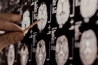




According to University of Iowa researchers Vincent A. Magnotta and colleagues, any neuroscientist with an MRI scanner could soon be able to measure the acidity (pH) of the human brain in great detail: Detecting activity-evoked pH changes in human brain.
If it works out, it would open up a whole new dimension of neuroimaging - and might be able to answer some of the biggest questions in the field.
The method relies on measuring T1 relaxation in the rotating frame (T1?). Essentially, it's about the rate at which protons are swapped between water molecules and proteins. That rate is known to depend on pH.
Anyway. It certainly looks impressive. Using a standard 3 Tesla MRI scanner, they were able to image the whole brain once every 6.6 seconds - only slightly slower than conventional fMRI measurements of brain activity, where 2 or 3 seconds is more usual. The spatial resolution was comparable to fMRI.
Here's how it did on some bottles of jelly -
Then they moved onto mouse brains (the differences are smaller here)...

And finally they scanned some people. They were able to detect the (very small) pH changes caused by hyperventilation, which raises pH, and breathing air enriched in carbon dioxide, which lowers it.

 Explore All Options
Lovely pictures I'm sure you agree, and it's a very clever methodology from a technical point of view. But what will it mean for neuroscience?
Well, for one thing, it might be able to help resolve some of the debates over what conventional fMRI is actually measuring. For example, some neuroscientists believe that many (seemingly) interesting fMRI results may actually be (at least partially) reflections of subtle changes in breathing rate. Measuring acidity, an indirect proxy for breathing, could start to answer such questions.
The main question though is, what are we going to call the new method? "T1? MRI"... not a terribly catchy name.
Maybe MRalkalI?
Magnotta, V., Heo, H., Dlouhy, B., Dahdaleh, N., Follmer, R., Thedens, D., Welsh, M., and Wemmie, J. (2012). Detecting activity-evoked pH changes in human brain Proceedings of the National Academy of Sciences DOI: 10.1073/pnas.1205902109